
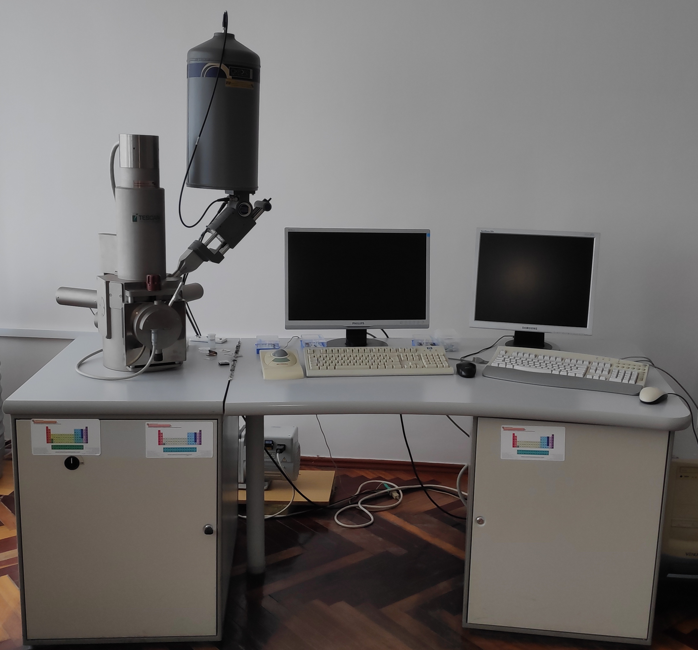
| Scanning Electron Microscope VEGA TS 5130 | |
|---|---|
| Scanning Electron Microscopy (SEM) uses a focused electron beam to scan small areas of solid samples. Secondary electrons are emitted from the sample and are collected to create an area map of the secondary emissions. Since the intensity of secondary emission is very dependent on local morphology, the area map is a magnified image of the sample. Spatial resolution is as high as 1 nanometer for some instruments, but 4 nm is typical for most. Magnification factors can exceed 500,000. Backscattered electrons (BSE) and characteristic X-rays are also generated by the scanning beam and many instruments can utilize these signals for compositional analysis of microscopically small portions of the sample. | |
|
Technique Advantages: Specifications: |
 |